38 heart structure and labels
The Anatomy of the Heart, Its Structures, and Functions Heart Anatomy The heart is made up of four chambers: Atria: Upper two chambers of the heart. Ventricles: Lower two chambers of the heart. Heart Wall The heart wall consists of three layers: Epicardium: The outer layer of the wall of the heart. Myocardium: The muscular middle layer of the wall of the heart. Endocardium: The inner layer of the heart. Heart Anatomy Labeling Game - PurposeGames.com This is an online quiz called Heart Anatomy Labeling Game. There is a printable worksheet available for download here so you can take the quiz with pen and paper. Your Skills & Rank. Total Points. 0. Get started! Today's Rank--0. Today 's Points. One of us! Game Points. 19. You need to get 100% to score the 19 points available.
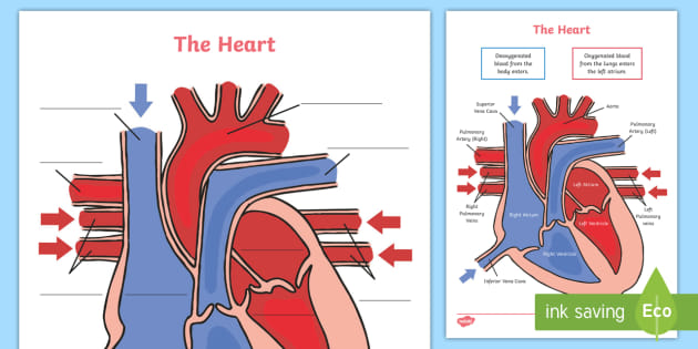
Heart Anatomy: Labeled Diagram, Structures, Function, and Blood Flow There are 4 chambers, labeled 1-4 on the diagram below. To help simplify things, we can convert the heart into a square. We will then divide that square into 4 different boxes which will represent the 4 chambers of the heart. The boxes are numbered to correlate with the labeled chambers on the cartoon diagram.

Heart structure and labels
webaim.org › standards › wcagWebAIM's WCAG 2 Checklist Feb 26, 2021 · 2.4.6 Headings and Labels (Level AA) Page headings and labels for form and interactive controls are informative. Avoid duplicating heading (e.g., "More Details") or label text (e.g., "First Name") unless the structure provides adequate differentiation between them. 2.4.7 Focus Visible (Level AA) Heart: illustrated anatomy - e-Anatomy - IMAIOS This interactive atlas of human heart anatomy is based on medical illustrations and cadaver photography. The user can show or hide the anatomical labels which provide a useful tool to create illustrations perfectly adapted for teaching. Anatomy of the heart: anatomical illustrations and structures, 3D model and photographs of dissection. Label Heart Anatomy Diagram Printout - EnchantedLearning.com Every day, the heart pumps about 2,000 gallons (7,600 liters) of blood, beating about 100,000 times. Label the heart anatomy diagram below using the heart glossary. Note: On the diagram, the right side of the heart appears on the left side of the picture (and vice versa) because you are looking at the heart from the front. Enchanted Learning Search
Heart structure and labels. Heart Diagram for Kids - Bodytomy The heart wall consists of three layers known as epicardium, myocardium and endocardium. Epicardium is the outer layer, myocardium is the middle layer, and endocardium is the innermost layer. Myocardium makes up for the bulk of the heart and is responsible for the pumping and beating of the heart. Atria and Ventricles Heart: Anatomy and Function - Cleveland Clinic A layer of muscular tissue called the septum divides your heart walls into the left and right sides. Your heart walls have three layers: Endocardium: Inner layer. Myocardium: Muscular middle layer. Epicardium: Protective outer layer. The epicardium is one layer of your pericardium. The pericardium is a protective sac that covers your entire heart. Diagrams, quizzes and worksheets of the heart | Kenhub Labeled heart diagrams Take a look at our labeled heart diagrams (see below) to get an overview of all of the parts of the heart. Once you're feeling confident, you can test yourself using the unlabeled diagrams of the parts of the heart below. Labeled heart diagram showing the heart from anterior Unlabeled heart diagrams (free download!) A Labeled Diagram of the Human Heart You Really Need to See The human heart, comprises four chambers: right atrium, left atrium, right ventricle and left ventricle. The two upper chambers are called the left and the right atria, and the two lower chambers are known as the left and the right ventricles. The two atria and ventricles are separated from each other by a muscle wall called 'septum'.
Human Heart Diagram Labeled | Science Trends List Of Heart Structures Heart Chambers Ventricles - The bottom two heart chambers. Atra - The upper two heart chambers. Wall Of The Heart Sinoatrial Node - A collection of tissue that releases electrical impulses and defines the rate of contraction for the heart. Atrioventricular Bundle - The fibers which transmit cardiac impulses. Human Heart (Anatomy): Diagram, Function, Chambers, Location in Body The right ventricle receives blood from the right atrium and pumps it to the lungs, where it is loaded with oxygen. The left atrium receives oxygenated blood from the lungs and pumps it to the left... How the Heart Works - What the Heart Looks Like | NHLBI, NIH The heart is made of three layers of tissue. Endocardium is the thin inner lining of the heart chambers and also forms the surface of the valves.; Myocardium is the thick middle layer of muscle that allows your heart chambers to contract and relax to pump blood to your body.; Pericardium is the sac that surrounds your heart. Made of thin layers of tissue, it holds the heart in place and ... byjus.com › biology › human-heartHuman Heart - Anatomy, Functions and Facts about Heart Following are the main functions of the heart: One of the primary functions of the human heart is to pump blood throughout the body. Blood delivers oxygen, hormones, glucose and other components to various parts of the body, including the human heart. The heart also ensures that adequate blood pressure is maintained in the body.
heart | Structure, Function, Diagram, Anatomy, & Facts | Britannica heart, organ that serves as a pump to circulate the blood. It may be a straight tube, as in spiders and annelid worms, or a somewhat more elaborate structure with one or more receiving chambers (atria) and a main pumping chamber (ventricle), as in mollusks. In fishes the heart is a folded tube, with three or four enlarged areas that correspond to the chambers in the mammalian heart. Heart anatomy: Structure, valves, coronary vessels | Kenhub Heart anatomy. The heart has five surfaces: base (posterior), diaphragmatic (inferior), sternocostal (anterior), and left and right pulmonary surfaces. It also has several margins: right, left, superior, and inferior: The right margin is the small section of the right atrium that extends between the superior and inferior vena cava . | The Heart Foundation The Heart Foundation saves lives and improves health through funding world-class cardiovascular research, guidelines for health professionals, informing the public and assisting people with cardiovascular disease Heart Diagram - 15+ Free Printable Word, Excel, EPS, PSD Template ... Label The Parts Of The Heart. depts.washington.edu | Having the heart diagram for studies or for scientific purpose has been made easy through this template. It shows a heart picture with all its parts labeled to make it easy to study. ... While studying anatomy heart is a major part to be covered. To study this lot of templates are available ...
How to Draw the Internal Structure of the Heart (with Pictures) To draw the internal structure of a human heart, follow the steps below. Part 1 Finding a Diagram 1 To find a good diagram, go to Google Images, and type in "The Internal Structure of the Human Heart". Find an image that displays the entire heart, and click on it to enlarge it. 2 Find a piece of paper and something to draw with.
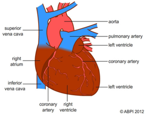
Structure of the Heart | The Franklin Institute The two largest veins that carry blood into the heart are the superior vena cava and the inferior vena cava. They are called "vena cava" because they are the "heart's veins." The superior is located near the top of the heart. The inferior is located beneath the superior. A wall called a septum, separates the right and left sides of the heart.
byjus.com › biology › diagram-of-heartHeart Diagram with Labels and Detailed Explanation - BYJUS Well-Labelled Diagram of Heart The heart is made up of four chambers: The upper two chambers of the heart are called auricles. The lower two chambers of the heart are called ventricles. The heart wall is made up of three layers: The outer layer of the heart wall is called epicardium. The middle layer of the heart wall is called myocardium.
Label the heart — Science Learning Hub In this interactive, you can label parts of the human heart. Drag and drop the text labels onto the boxes next to the diagram. Selecting or hovering over a box will highlight each area in the diagram. Right ventricle Right atrium Left atrium Pulmonary artery Left ventricle Pulmonary vein Semilunar valve Vena cava Aorta Download Exercise Tweet
Easy way to draw heart structure by 5 steps - YouTube My youtube channel : facebook page : way to draw hea...
Heart Blood Flow | Simple Anatomy Diagram, Cardiac Circulation ... - EZmed Diagram: Blood flow through the right side of the heart involving the following cardiac structures: superior vena cava (SVC), inferior vena cava (IVC), right atrium (RA), tricuspid valve (TV), right ventricle (RV), pulmonary valve (PV), and main pulmonary artery (PA). Trick to Remember the Right Side
› user › VideoJugVideojug - YouTube Welcome to Videojug! Here you'll find the best how-to videos around, from delicious, easy-to-follow recipes to beauty and fashion tips.
Heart Labels - Printable or Custom Printed Stickers | Avery.com Use our free specialty shape label templates to easily personalize your heart labels online. Customize one of our free designs or upload your own graphics and then choose the printing option that works best for you. Order your blank heart labels or custom printed heart labels and stickers online and get free shipping on orders of $50 more.

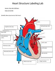
Lab Heart Structure Labeling. - Heart Structure Labeling Lab Name Hannah Kingsley Date Time 11 ...
› metal-hammerMetal Hammer | Louder 1 day ago · Five Finger Death Punch get up close and personal, only in the new issue of Metal Hammer. By Metal Hammer published 21 July 22 The star-packed new issue of Metal Hammer is out now, featuring Five Finger Death Punch, Judas Priest, Arch Enemy, Ross Robinson, Jinjer and more
Heart Diagram with Labels and Detailed Explanation The heart is located under the ribcage, between the lungs and above the diaphragm. It weighs about 10.5 ounces and is cone shaped in structure. It consists of the following parts: Heart Detailed Diagram Heart - Chambers There are four chambers of the heart . The upper two chambers are the auricles and the lower two are called ventricles.
The structure of the heart - Structure and function of the heart ... It is located in the middle of the chest and slightly towards the left. The heart is a large muscular pump and is divided into two halves - the right-hand side and the left-hand side. The...
Cross Section of the Heart Diagram & Function | Body Maps Cross-section. The chambers of the heart operate as a 'double-pump' system for the body's circulation. In coordination with valves, the chambers work to keep blood flowing in the proper ...
ods.od.nih.gov › factsheets › Calcium-ConsumerCalcium - Consumer Almost all calcium in the body is stored in bones and teeth, giving them structure and hardness. Your body needs calcium for muscles to move and for nerves to carry messages between your brain and every part of your body. Calcium also helps blood vessels move blood throughout your body and helps release hormones that affect many functions in ...
Structure of the Heart | SEER Training The human heart is a four-chambered muscular organ, shaped and sized roughly like a man's closed fist with two-thirds of the mass to the left of midline. The heart is enclosed in a pericardial sac that is lined with the parietal layers of a serous membrane. The visceral layer of the serous membrane forms the epicardium. Layers of the Heart Wall
Diagram of Human Heart and Blood Circulation in It A heart diagram labeled will provide plenty of information about the structure of your heart, including the wall of your heart. The wall of the heart has three different layers, such as the Myocardium, the Epicardium, and the Endocardium. Here's more about these three layers. Epicardium
Label Heart Anatomy Diagram Printout - EnchantedLearning.com Every day, the heart pumps about 2,000 gallons (7,600 liters) of blood, beating about 100,000 times. Label the heart anatomy diagram below using the heart glossary. Note: On the diagram, the right side of the heart appears on the left side of the picture (and vice versa) because you are looking at the heart from the front. Enchanted Learning Search
Heart: illustrated anatomy - e-Anatomy - IMAIOS This interactive atlas of human heart anatomy is based on medical illustrations and cadaver photography. The user can show or hide the anatomical labels which provide a useful tool to create illustrations perfectly adapted for teaching. Anatomy of the heart: anatomical illustrations and structures, 3D model and photographs of dissection.
webaim.org › standards › wcagWebAIM's WCAG 2 Checklist Feb 26, 2021 · 2.4.6 Headings and Labels (Level AA) Page headings and labels for form and interactive controls are informative. Avoid duplicating heading (e.g., "More Details") or label text (e.g., "First Name") unless the structure provides adequate differentiation between them. 2.4.7 Focus Visible (Level AA)


_pt.svg.png)


Post a Comment for "38 heart structure and labels"