39 microscope labels and definitions
Types of Microscopes - Laboratoryinfo.com It is an optical type of microscope enabling the viewer to see the sample in 3-dimensions. Its magnification power ranges from 10x to 80x. It is primarily used to inspect large specimens like fossils, rocks, coins, hair follicles, stamps, parts of flowers, and the likes. Image 19: A gemological microscope is perfect for examining gems and minerals. Simple Microscope - Diagram (Parts labelled), Principle, Formula and Uses A simple microscope consists of Optical parts Mechanical parts Labeled Diagram of simple microscope parts Optical parts The optical parts of a simple microscope include Lens Mirror Eyepiece Lens A simple microscope uses biconvex lens to magnify the image of a specimen under focus.
CFR - Code of Federal Regulations Title 21 29/03/2022 · The provisions of this section are not applicable to systems which are designed exclusively for microscopic examination of material, e.g., x-ray diffraction, spectroscopic, and electron microscope equipment or to systems for intentional exposure of humans to x-rays. (b) Definitions. As used in this section the following definitions apply:
Microscope labels and definitions
Electron Microscope- Definition, Principle, Types, Uses, Labeled Diagram An electron microscope is a microscope that uses a beam of accelerated electrons as a source of illumination. It is a special type of microscope having a high resolution of images, able to magnify objects in nanometres, which are formed by controlled use of electrons in a vacuum captured on a phosphorescent screen. Light Microscope (Theory) - Amrita Vishwa Vidyapeetham To study the various parts and working of a microscope. Microscope Microscope is an optical instrument that uses lens or combination of lens to produce magnified images that are too small to seen by unaided eye. Microscope provides the enlarged view that helps in examining and analyzing the image. Compound Microscope - Diagram (Parts labelled), Principle and Uses See: Labeled Diagram showing differences between compound and simple microscope parts Structural Components The three structural components include 1. Head This is the upper part of the microscope that houses the optical parts 2. Arm This part connects the head with the base and provides stability to the microscope.
Microscope labels and definitions. Microscope, Microscope Parts, Labeled Diagram, and Functions Microscopes magnify or enlarge small objects such as cells, microbes, bacteria, viruses, microorganisms etc. at a viewable scale for examination and analysis. Microscopes consist of one or more magnification lenses to enlarge the image of the microscopic objects placed in the focal plane. Compound Microscope- Definition, Labeled Diagram, Principle, Parts, Uses Therefore, a microscope can be understood as an instrument to observe tiny elements. The optical microscope often referred to as the light microscope, is a type of microscope that uses visible light and a system of lenses to magnify images of small subjects. There are two basic types of optical microscopes: Simple microscopes Compound microscopes microscope | Types, Parts, History, Diagram, & Facts | Britannica Optical microscopes can be simple, consisting of a single lens, or compound, consisting of several optical components in line. The hand magnifying glass can magnify about 3 to 20×. Single-lensed simple microscopes can magnify up to 300×—and are capable of revealing bacteria —while compound microscopes can magnify up to 2,000×. Light Microscope- Definition, Principle, Types, Parts, Labeled Diagram ... A light microscope is a biology laboratory instrument or tool, that uses visible light to detect and magnify very small objects and enlarge them. They use lenses to focus light on the specimen, magnifying it thus producing an image. The specimen is normally placed close to the microscopic lens.
What is an Electron Microscope? - Definition, Types & Uses An electron microscope allows us to see at these small scales. Electron microscopes work by using an electron beam instead of visible light and an electron detector instead of our eyes. An ... List of Laboratory Safety Symbols and Their Meanings 1. General Warning 2. Biohazard 3. Flammable Material Hazard 4. Explosive Material Hazard 5. Electrical Hazard 6. High Voltage Hazard 7. Toxic Material Hazard 8. Ionizing Radiation Hazard 9. Non-Ionizing Radiation Hazard 10. Low Temperature Hazard 11. Hot Surface Hazard 12. UV Light hazard 13. Oxidizing Material Hazards 14. Testes: Anatomy, definition and diagram | Kenhub Testis. 1/5. The testes (testicles) are male reproductive glands found in a saccular extension of the anterior abdominal wall called the scrotum. They are in ovoid shape, sized four to six centimeters in length. Testes develop retroperitoneally on the posterior abdominal wall and descend to scrotum before birth. Simple Microscope- Definition, Principle, Magnification, Parts ... Simple Microscope Definition A simple microscope is one that uses a single lens for magnification, such as a magnifying glass while a compound microscope uses several lenses to enhance the magnification of an object. It uses a lens to enlarge an object through angular magnification alone, giving the viewer an erect enlarged virtual image.
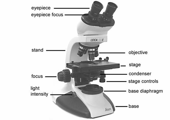
Parts of a Compound Microscope and Their Functions The main parts of compound microscope are the condenser lens, the objective lens, and the eyepiece lens, and these instruments are referred to as compound microscopes. Each of these components is made up of microscope lens combinations that are required to produce magnified images with minimal artefacts and aberrations. Structure of Microscope Light Microscope: Definition, Uses & Parts - Study.com A light microscope uses focused light and lenses to magnify a specimen, usually a cell. In this way, a light microscope is much like a telescope, except that instead of the object being very large ... Microscope Parts | A Guide on their Location and Function The image of a compound microscope with labeled parts. Eyepiece It is the part that you encounter when viewing an object in the microscope from the top. This is the first lens that helps to magnify the image. Based on the magnification power, the lens can be of 5X, 10X, 15X, or more. Using A Microscope 101: Important Microscope Parts & Functions Bring a microscope to class and point to the different parts. Have the students label a diagram of a microscope. During activities that involve microscopes, make sure you ask each student to name a few parts. Ask students to explain how they would prepare a slide or transport a microscope. Make sure they use the correct names for the different ...
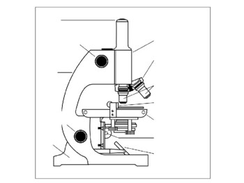
32 Picture Of A Microscope To Label - Labels 2021
Microscope Types (with labeled diagrams) and Functions A compound microscope: Is used to view samples that are not visible to the naked eye. Uses two types of lenses - Objective and ocular lenses. Has a higher level of magnification - Typically up to 2000x. Is used in hospitals and forensic labs by scientists, biologists and researchers to study micro organisms. Compound microscope labeled diagram.

Definitions Label Clip Art at Clker.com - vector clip art online, royalty free & public domain
Parts of a microscope with functions and labeled diagram Microscope Definition Microscopes are instruments that are used in science laboratories to visualize very minute objects such as cells, and microorganisms, giving a contrasting image that is magnified. Microscopes are made up of lenses for magnification, each with its own magnification powers.

labels of a compound microscope compound microscope worksheets 23 - Top Label Maker
Compound Microscope - Types, Parts, Diagram, Functions and Uses Compound microscope - It is an optical instrument consists of two convex lenses of short focal lengths primarily used for observing a highly magnified image of minute objects. Lenses Simple microscope - It has a convex lens. It uses only one lens to magnify objects. An example of a simple microscope is a magnifying glass.

Demassed : Entertainment Without An Entertainment Industry
20 most common lab equipment names, pictures and their uses A light microscope placed on a white surface. Photo: pexels.com, @Artem Podrez Source: UGC. A microscope is a popular lab apparatus used to observe things that are too tiny to be observed by the naked human eye. There are many different types of microscopes. A light microscope uses lights and a series of magnifying lenses to observe a tiny ...

34 Label A Compound Microscope - Labels Information List
Microscope Quiz: How Much You Know About Microscope Parts ... - ProProfs The microscope has been used in science to understand elements, diseases, and cells. You must have used a microscope back in high school in the biology lab. Do you believe you understood how to use it? Take up the test and see. Questions and Answers 1. Arm: A. You look through to see the specimen. B. Holds the slide in place. C.

35 Label Of Compound Microscope - Labels Information List
Microscope- Definition, Parts, Functions, Types, Diagram, Uses A microscope is an optical instrument having one or more lenses system which is used to get a clear magnified image of minute objects or structures that can't be viewed by the naked eyes. Derived from Greek words "mikrós " meaning "small" and "skópéō" meaning "look at " . They are devices used to observe the detailed structure of small objects.
Microscopes Applied
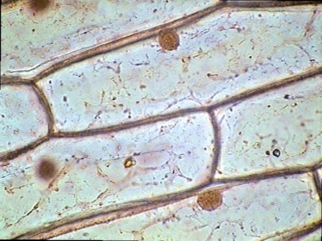
Different Size, Shape and Arrangement of Bacterial Cells Size of Bacterial Cell. The average diameter of spherical bacteria is 0.5-2.0 µm. For rod-shaped or filamentous bacteria, length is 1-10 µm and diameter is 0.25-1 .0 µm. E. coli , a bacillus of about average size is 1.1 to 1.5 µm wide by 2.0 to 6.0 µm long. Spirochaetes occasionally reach 500 µm in length and the cyanobacterium.

Lab Manual Exercise # 1
What is a Microscope Condenser? (with pictures) - Info Bloom Microscope condensers allow for light, contrast and clarity when viewing an object. The condenser consists of a lens or set of lenses mounted directly under the stage. The intensity of the light can be adjusted by moving the microscope condenser closer to or further away from the stage, and the width of the beam can be adjusted by making the ...

Biology label part of microscope
Compound Microscope Parts, Diagram Definition, Application, Working ... Definition of a Compound Microscope and uses. parts of a compound microscope and application. compound microscope labeled diagram. MN . Generic selectors. ... For future reference, adhesive labels are stuck to the base and sides of the microscope. (iii) Use. After calibrating the eyepiece scales for all objective lenses, the microscope can be ...

Label the microscope please don't label another microscope and send the picture. - Brainly.in
Parts of the Microscope with Labeling (also Free Printouts) Parts of the Microscope with Labeling (also Free Printouts) A microscope is one of the invaluable tools in the laboratory setting. It is used to observe things that cannot be seen by the naked eye. Table of Contents 1. Eyepiece 2. Body tube/Head 3. Turret/Nose piece 4. Objective lenses 5. Knobs (fine and coarse) 6. Stage and stage clips 7. Aperture
Microscope labelling 11 - Teaching resources
E-Learning – AOAC India Bright Field Microscope, Dark Field Microscope, Phase Contrast Microscope, Fluorescence Microscope, Confocal microscopy, Scanning and Transmission Electron Microscope and applications: 17th June 2019, 2019 11.30-12.30 pm: Watch Video: 50 : Nuclear magnetic resonance (NMR) – Part 1 DR. CHANDRASHEKHAR MR. RAGHAV MAVINKURVE, BRUKER
:max_bytes(150000):strip_icc()/microscopestudy-58b978755f9b58af5c495ac7.png)
Learn About Microscopes With Fun, Free Printables
Simple Microscope - Parts, Functions, Diagram and Labelling Simple Microscope - Parts, Functions, Diagram and Labelling A microscope is one of the commonly used equipment in a laboratory setting. A microscope is an optical instrument used to magnify an image of a tiny object; objects that are not visible to the human eyes. Table of Contents The common types of microscopes are: What is a Simple microscope?
چگونه از میکروسکوپ نوری استفاده کنیم؟
Compound Microscope - Diagram (Parts labelled), Principle and Uses See: Labeled Diagram showing differences between compound and simple microscope parts Structural Components The three structural components include 1. Head This is the upper part of the microscope that houses the optical parts 2. Arm This part connects the head with the base and provides stability to the microscope.

1 set Leica DM6000 CS / TCS SP5 Laser inverted confocal microscope Win10 | eBay
Light Microscope (Theory) - Amrita Vishwa Vidyapeetham To study the various parts and working of a microscope. Microscope Microscope is an optical instrument that uses lens or combination of lens to produce magnified images that are too small to seen by unaided eye. Microscope provides the enlarged view that helps in examining and analyzing the image.

Microscope Gossary of Terms | The Microscope Depot
Electron Microscope- Definition, Principle, Types, Uses, Labeled Diagram An electron microscope is a microscope that uses a beam of accelerated electrons as a source of illumination. It is a special type of microscope having a high resolution of images, able to magnify objects in nanometres, which are formed by controlled use of electrons in a vacuum captured on a phosphorescent screen.
31 Label The Indicated Parts Of The Microscope - Labels For Your Ideas

Introductory Biology
8.1 & 8.2 Math and Science: Onion Skin Lab
Post a Comment for "39 microscope labels and definitions"